


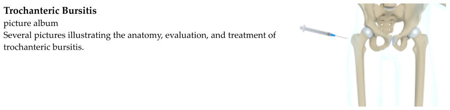
Trochanteric Bursitis
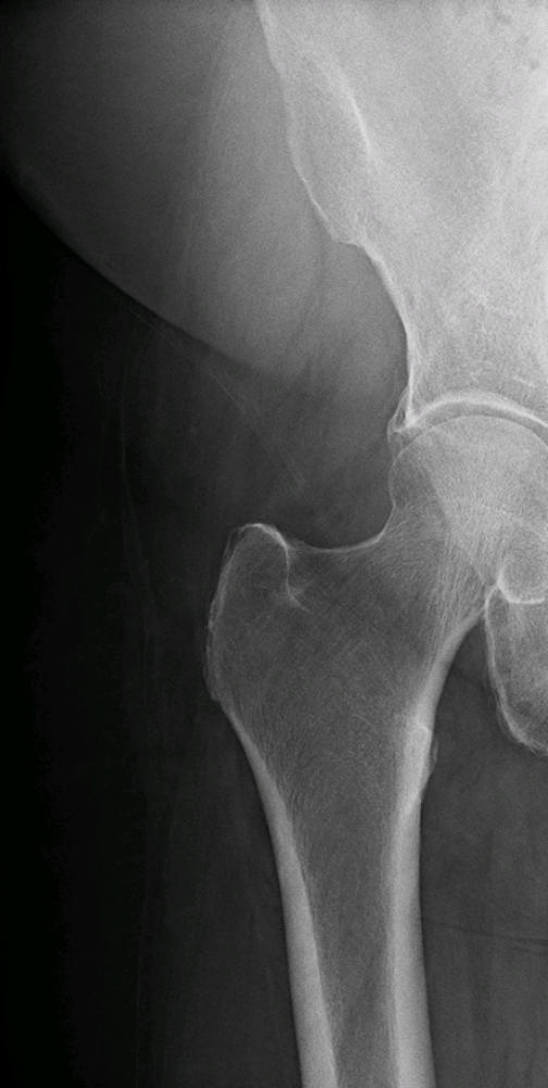
The greater trochanter is a bony prominence on the femur (the thigh bone). It can be felt on the side of the hip.
Several muscles attach to or pass over the femur. Movement creates friction as the muscles and tendons rub
against the bone. A bursa is a thin sack of fluid that lies between the bone and the soft tissue structures that helps
to limit friction and prevent inflammation. The trochanteric bursa lies between the greater trochanter of the
femur (the point of the hip) and a long tendon known as the iliotibial band.
Trochanteric bursitis is when the bursae, muscles, and tendons around the greater trochanter become inflamed.
Inflammation may occur with a traumatic blow to the hip. It may also occur as the muscles and tendons rub back
and forth against the bone and bursae. Running, especially on uneven ground, a leg length discrepancy (one leg is
slightly shorter than the other), and anatomical differences can predispose people to developing trochanteric
bursitis.
Trochanteric bursitis is diagnosed based on a history and physical exam. Patients may report a history of a blow to
the hip, perhaps as the result of a fall or while playing contact sports. Runners are also predisposed, especially if
they run on a slanted surface. People who have had hip surgery can develop hip bursitis as well.
During the physical exam, the hip is tender over the greater trochanter. People with a leg length discrepancy are
more likely to develop trochanteric bursitis.
X-rays may be obtained to look for other causes of hip pain such as a fracture of the greater trochanter.
Trochanteric bursitis is almost always treated without surgery. Treatment includes non- steroidal anti-
inflammatory drugs (NSAIDs) such as ibuprofen, correction of any leg length discrepancy, stretching exercises,
ice, and activity modification. Steroid injections (cortisone shots) into the trochanteric bursae can provide relief.
Physical therapy may also be beneficial.
Physical therapy involves stretching the iliotibial band and other muscles and tendons surrounding the hip. Other
modalities such as phonophoresis (ultrasound and application of steroid cream) may be utilized by the physical
therapist as well.
If non-surgical treatment is not effective, surgical release of the iliotibial band and removal of the bursae has been
successful in relieving symptoms.