


Scaphoid Fracture
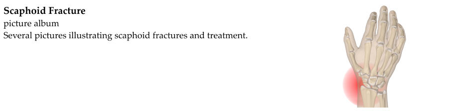
The carpals are the small bones of the wrist. One of the carpals, located near the base of the thumb, is known as the
scaphoid.
Fracture is another term for broken bone. There is no difference between a fracture and a broken bone. And so a
scaphoid fracture is a broken bone in the wrist. The scaphoid is by far the most commonly broken of the carpal
bones.
Scaphoid fractures usually occur as the result of a fall onto an outstretched hand. They are more common in people
under the age of 70. People older than 70 usually break the distal radius rather than the scaphoid when they fall.
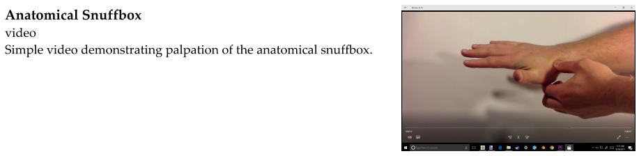
People with scaphoid fractures are usually very tender over an area of the wrist known as the anatomical snuffbox.
This is a little depression between two tendons at the base of the thumb.
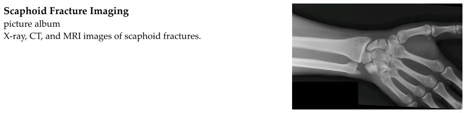
Scaphoid fractures are usually visible on x-rays. The x-rays are used to evaluate the fracture and form a treatment
plan. Sometimes, the scaphoid fracture is a crack that cannot be seen on the initial x-rays. This is known as an
occult fracture. If a scaphoid fracture is suspected but not visible on x-rays, a couple of options are available. First,
the wrist can be placed in a cast and treated as if it was known to be broken. The cast is removed in 10-14 days and
new x-rays obtained. The fracture may be more visible after this time. The second option is to obtain another
imaging study such as an MRI, CT scan, or bone scan. These studies can be helpful in identifying or ruling out
fractures that were not visible on regular x-rays.
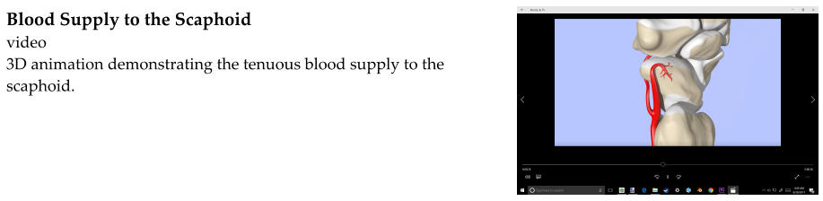
Once the fracture has been identified, it is important to treat it correctly. The scaphoid has a reputation for not
healing as well as most other bones. This is because of the way that blood is supplied to the scaphoid. Blood is
essential for healing because it carries nutrients to the bone that allow it to remain healthy and heal. Blood is
supplied to the scaphoid by small branches from the radial artery. These small branches enter at the end of the
scaphoid nearest the thumb. The blood then has to travel through the bone tissue to the other end of the scaphoid.
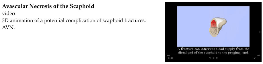
A fracture can interrupt this blood flow. If the fracture occurs at the end with poor blood supply, it will take longer
to heal. It is also at a higher risk of non-union, meaning that it never heals.
Another complication that can occur with scaphoid fractures is avascular necrosis (AVN). This can occur if the
fracture is displaced enough to interrupt the blood supply from one end of the bone to the other. “Displaced”
means that the bone fragments are pulled apart. If blood supply to one end of the bone is interrupted, that part of
the bone is deprived of nutrients that it needs and can die.
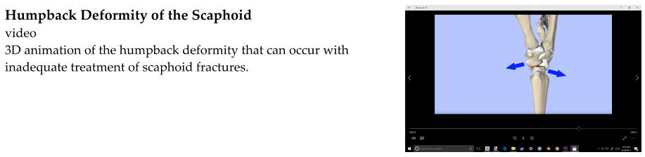
A third complication that can occur with scaphoid fractures is carpal instability which can result in osteoarthritis,
chronic pain, and weakness. There are many ligaments that hold the carpal bones together. They are able to move
in concert to allow for strong, smooth motion at the wrist. If the scaphoid does not heal correctly (malunion), it
can disrupt the mechanics of the wrist.
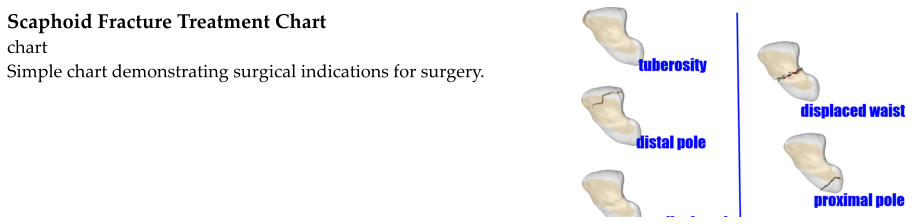
Whether to treat a scaphoid fracture with casting or with surgery is usually decided with the help of an orthopaedic
surgeon. Nondisplaced fractures of the distal end or waist of the scaphoid are usually treated with casting.
Displaced fractures are usually treated with surgery. Because of slow healing and risk of non-union, fractures of the
proximal scaphoid are often treated with surgery as well.
If non-operative treatment is chosen, the wrist is placed in a long-arm thumb spica cast. The long arm cast can
be replaced with a short arm thumb spica cast after six weeks. Depending on where the fracture is located
(remember that fractures at the proximal end heal more slowly), the short arm cast is left in place for six to ten
weeks. Because the wrist is immobilized for a long period of time, it will be stiff and weak after the cast is removed.
Physical therapy can help to improve range of motion and strength.
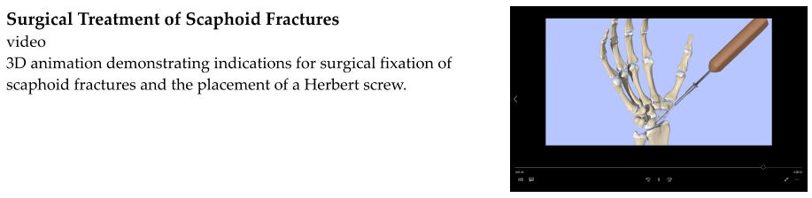
Surgical treatment involves using a screw to compress the fragments together. This gives the scaphoid a better
chance of healing correctly and more quickly. A thumb spica cast may still be applied after surgery, but does not
have to be applied for as long as a cast would if the fracture were treated non-operatively. Physical therapy may be
necessary as the fracture heals.