


Rotator Cuff Syndrome


The rotator cuff is a group of four muscles in the shoulder that help to elevate and rotate the arm. They originate on
the scapula, the shoulder blade, and attach to the head of the humerus, the upper arm bone. Tendons are strong,
fibrous bands that attach muscles to bones. The rotator cuff muscles become tendons near their attachments to the
humerus.
Rotator cuff syndrome is a spectrum of injuries to the rotator cuff ranging from tendinitis, or inflammation of the
rotator cuff tendons, to full-thickness tears. It may be caused by shoulder impingement. This occurs when the space
where the rotator cuff tendons pass between the head of the humerus and the part of the scapula known as the
acromion becomes too tight and pinches the rotator cuff. It may also be caused by an injury or overuse such as with
overhead throwing athletes.
Symptoms of rotator cuff syndrome include shoulder pain with attempts to raise the arm overhead or reach behind
the back. Over time, the shoulder can become very weak. Different physical exam maneuvers have been developed
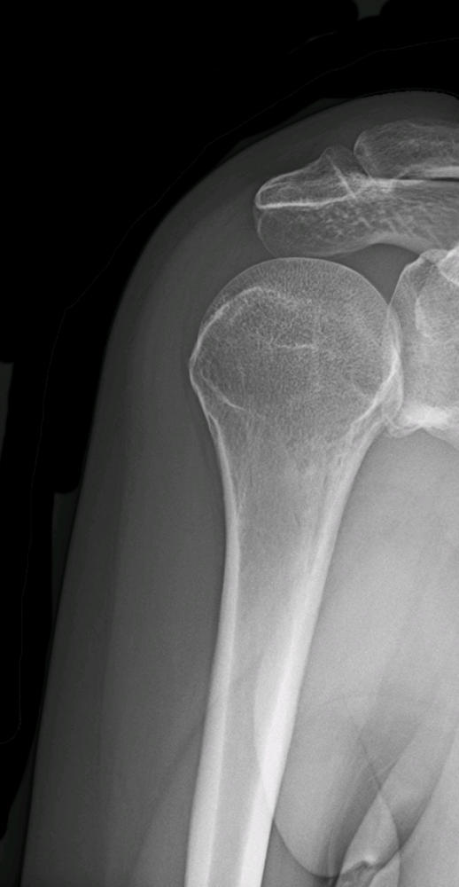
to evaluate the different rotator cuff muscles and distinguish them from other shoulder problems. X-rays may
demonstrate bone spurs or a hooked acromion that is impinging upon the rotator cuff. They may also demonstrate
cystic changes in the humeral head common with chronic rotator cuff tears. The rotator cuff itself is not visible on x-
rays. If a full-thickness tear is suspected, an MRI may be obtained. The rotator cuff muscles and tendons can be
visualized on an MRI and impingement, tendinitis, bursitis, and tears can be evaluated.
Treatment for rotator cuff syndrome is usually non-surgical. Anti-inflammatory medications can reduce
inflammation and relieve pain. Physical therapy and activity modification can strengthen the rotator cuff and
surrounding muscles. A steroid injection into the subacromial space can also be effective.
If there is a full-thickness tear of the rotator cuff, there is less potential to heal without surgery. Surgery is usually
done arthroscopically, meaning that a small camera and instruments are inserted into the shoulder through small,
keyhole incisions. The damaged portion of the rotator cuff is debrided. The healthy portion is anchored down to the
bone using anchors and suture.
Recovery after a rotator cuff repair usually takes about 3 to 4 months. For the first several weeks, the arm is placed
in a sling to avoid putting too much stress across the repaired tendon. Once the tendon has healed down to the bone,
physical therapy can help to restore range-of-motion and strength.
Anatomy of the Rotator Cuff
The shoulder joint is where the humerus, the upper arm bone, meets the scapula, the shoulder blade. The head of
the humerus forms a ball that sits in a socket formed by the scapula. This is known as the glenoid. Above this
glenohumeral joint is a bony process -- also part of the scapula -- called the acromion. The rotator cuff muscles
originate on the scapula.
The subscapularis sits on the front of the scapula in the subscapular fossa. It passes across the front of the shoulder
and attaches to the lesser tubercle of the humerus. As the subscapularis contracts, it pulls the humerus inward and
downward, internally rotating the arm. And so the subscapularis is important in reaching behind the back.
The supraspinatus sits on the top of the scapula in the suprascapular fossa. It then passes underneath the acromion
and over the top of the humeral head. It attaches to the greater tubercle of the humeral head. As the supraspinatus
contracts, it pulls the humerus toward the center of the body. This helps to elevate the arm up to the side, a motion
known as abduction. The larger deltoid muscle is also involved in shoulder abduction, but the supraspinatus is
especially important during the first 15°.
The infraspinatus originates on the back of the scapula in the infraspinous fossa and attaches to the greater tubercle
of the humerus. As the infraspinatus contracts, it helps to externally rotate the arm.
The teres minor originates on the side of the scapula, the lateral border, and attaches to the lower part of the greater
tubercle of the humerus. It also contributes to external rotation.
All of the rotator cuff muscles help to stabilize the shoulder by holding the humeral head in the correct position in
the glenoid.
Between the rotator cuff and the acromion is the subacromial bursa. This is often described as a sack of fluid. Its
purpose is to decrease the amount of friction between the rotator cuff and the acromion.
Shoulder Impingement Syndrome and Bursitis
The rotator cuff muscles attach the humerus to the scapula and help to elevate and rotate the arm. The rotator cuff
muscle that originates at the top of the scapula is called the supraspinatus. The supraspinatus passes just below a
bony process on the scapula known as the acromion. The space between the acromion and the humeral head is
known as the subacromial space. A bursa, soft, fluid-filled tissue, sits on top of the rotator cuff and helps to limit
friction between the supraspinatus and the acromion.
When the subacromial space becomes too tight, it can squeeze and impinge upon the rotator cuff. This causes
friction and inflammation. This is known as shoulder impingement syndrome. If the subacromial bursa becomes
irritated and inflamed, the condition can also be referred to as shoulder bursitis.
Anything that causes decreased subacromial space can lead to impingement syndrome. Some people have a hooked
acromion. The hooked portion of the acromion points downward into the subacromial space and the rotator cuff.
Many people develop arthritis in the acromioclavicular joint, where the acromion meets the clavicle, as they age.
This can cause bone spurs and hypertrophy of the AC joint which narrows the subacromial space.
Shoulder impingement syndrome usually responds to conservative treatment. Non-steroid anti-inflammatory drugs
such as ibuprofen can help decrease pain and swelling. Physical therapy can stretch and strengthen the associated
muscles. A steroid injection given into the subacromial space can decrease inflammation as well.
If shoulder impingement syndrome does not improve with non-operative treatment, an MRI is usually obtained to
rule out other causes of shoulder pain such as a torn rotator cuff.
Shoulder impingement syndrome can be addressed surgically by performing a subacromial decompression. This is
typically done arthroscopically. The bottom of the acromion is shaved away to open up the subacromial space. The
inflamed bursa can also be excised or debrided.