


Olecranon Fracture
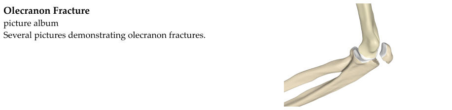
The part of the ulna that forms a hinge with the humerus is called the olecranon. The smooth, round part of the
humerus the fits to the olecranon is known as the trochlea.
Olecranon fractures often occur as the result of a fall or other direct trauma to the point of the elbow. They may
also occur with a fall on an outstretched arm accompanied by a strong contraction of the triceps muscle.
Olecranon fractures cause elbow pain, especially at the back of the elbow. The person may be unable to extend
(straighten out) the elbow. There may be a divot just below the tip of the elbow. Elbow movement is painful.
Swelling and bruising are often present.
Pain/tenderness at the olecranon
Pain with elbow motion
Possible deformity
Possible inability to actively straighten the elbow
Diagnosis is based on:
History - fall or blow to the elbow resulting in elbow pain.
Physical exam - tenderness over the olecranon with or without deformity or inability to extend the elbow.
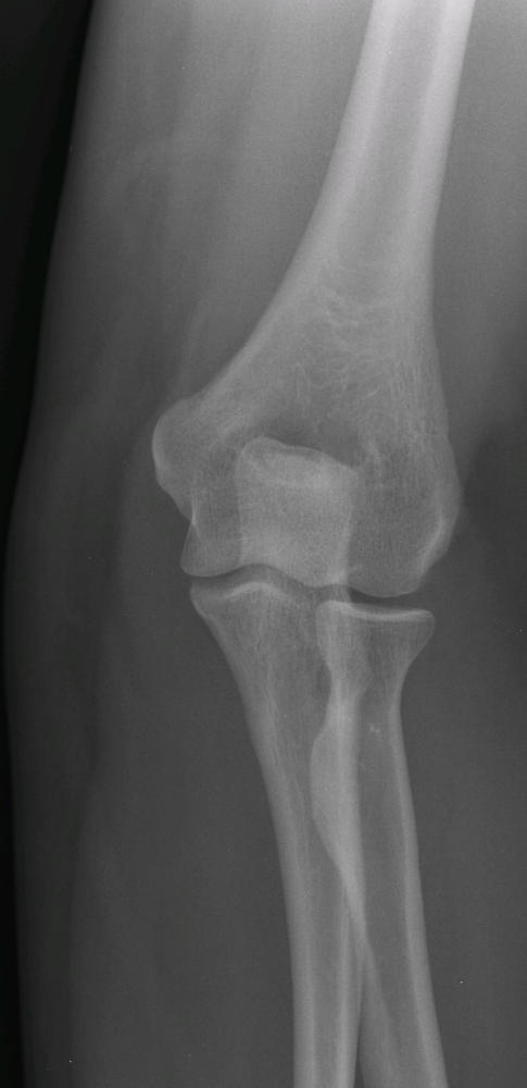
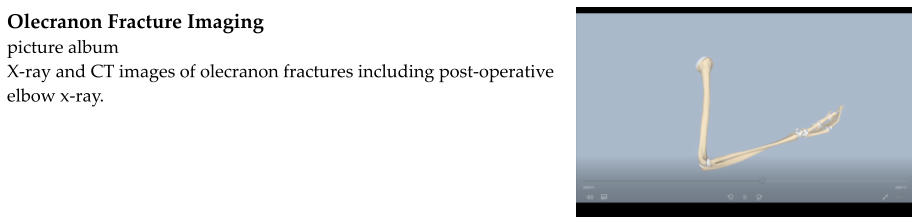
X-rays
CT scan - a CT scan gives a more detailed, 3- dimensional view of the fracture. A CT scan is not always necessary,
but can be helpful in evaluating the fracture if surgery is being considered.
A CT scan can provide a more detailed, 3-dimensional view.
Treatment for olecranon fractures depends on whether the olecranon is displaced and on whether the person with
the fracture is healthy and needs a well-functioning elbow.
A displaced olecranon fracture can result in damage to the cartilage that pads the elbow joint. It can also change
the mechanics of the elbow so it does not bend correctly. Surgery involves making an incision over the olecranon to
expose the fracture. The broken fragment is pushed back into place (reduced) and fixed in place with hardware
such as a plate and screws. This is known as open reduction-internal fixation (ORIF).
Nondisplaced fractures have the potential to heal without surgery.
Long arm cast with elbow bent to about 45-90°
Ice, elevation
Rest (no use of the arm)
Protected range-of-motion exercises after 3 to 6 weeks X-rays are obtained every 1-2 weeks to ensure that the
fracture is healing well. When there is evidence of solid healing, activity can be gradually increased.