


Clavicle Fracture
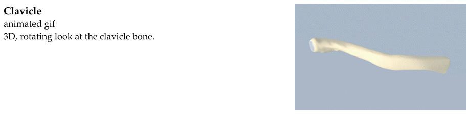
The clavicle is commonly known as the “collar bone.” A fracture is a broken bone.
And so a clavicle fracture is a broken collar bone.
Clavicle fractures usually occur as the result of a fall onto the shoulder. They may also
result from a direct blow to the clavicle or a fall onto an outstretched hand. They may
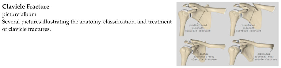
be nondisplaced (just cracked) or displaced (the broken fragments are pulled apart).
Most clavicle fractures occur near the middle of the shaft. Some fracture occur one of
the ends of the clavicle.
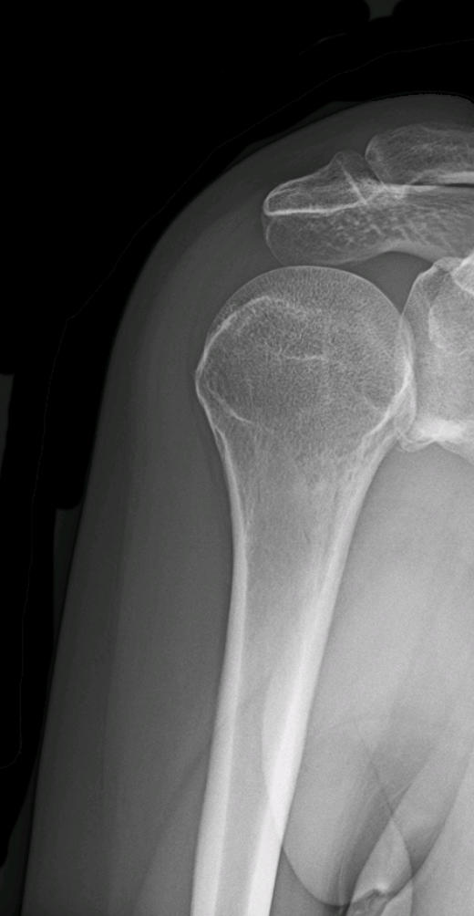
Clavicle fractures are diagnosed based on a history, physical exam, and x-rays.
Patients usually report a fall or a blow to the shoulder. They complain of pain and
sometimes a deformity and swelling over the collar bone.
Upon physical exam, the shoulder may be bruised and swollen over the clavicle. The
broken bone will, of course, be tender to push on. More severe clavicle fractures may
result in an obvious deformity. When this is the case, it is important to ensure that
the sharp fragment is not threatening to poke through the skin. This is known as
“tenting” of the skin.
X-rays are important not only to confirm that the clavicle is broken, but to evaluate the
fracture. Fractures may be non-displaced (just a crack) or displaced. They may
involve the cartilage in the joint (intra-articular). The fragments may overlap one
another, resulting in shortening of the clavicle.
Non-displaced clavicle fractures and clavicle shaft fractures that have less than 1-2 cm
of displacement or shortening can be treated without surgery. The arm is placed in a
sling, shoulder immobilizer, or figure-of-eight brace. The shoulder is immobilized in
this way for 4-6 weeks as the bone heals. Strenuous activity is not permitted until
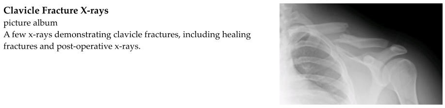
significant healing has occured. Follow-up x-rays are obtained to evaluate healing
and will demonstrate callus formation at the fracture site. Range-of-motion exercises
with the elbow and wrist are encouraged to prevent stiffness.
If the clavicle fracture is too displaced or shortened (greater than 1-2 cm), if the broken
fragments have poked through the skin or threaten to break through the skin, or if
there is a displaced fracture at the end of the clavicle, surgery may be indicated.
Surgery involved pushing the broken fragments back into placed and fixing them
together with hardware such as a plate and screws. This is called open reduction-
internal fixation or ORIF.