


Calcaneus Fracture


The calcaneus is one of the tarsal bones of the hindfoot. It is commonly referred to as the heel bone.
A calcaneus fracture, in simple terms, is a broken heel bone.
Calcaneus fractures are often the result of high-energy injuries such as a fall from a height or a motor
vehicle accident.
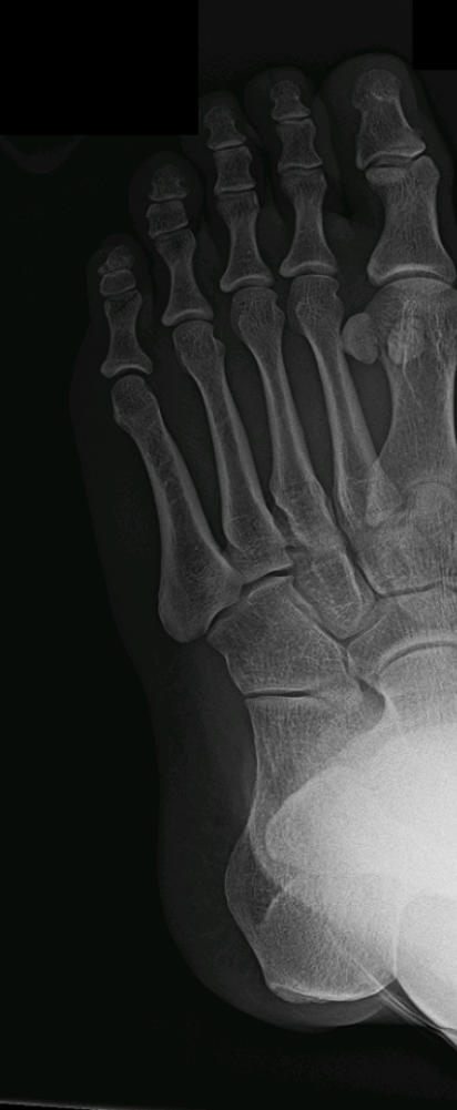
X-rays:
•
AP, lateral of foot
•
AP, lateral, oblique of ankle
•
Harris view (axial heel view)
•
Consider x-rays of the lumbar spine due to the high incidence of associated vertebral fractures
CT scan:
If the fracture is intra-articular (involves the joint surface), a CT scan should be obtained.
MRI:
If no fracture is identified but a stress fracture is suspected, an MRI may confirm this.
Classification:
•
Extra-articular
o
Anterior process (often missed; “sprain fractures”)
o
Tuberosity
▪
Open beak
▪
Avulsion at insertion of Achilles tendon
o
Sustentaculum tali
o
Body - not involving the subtalar joint
o
Medial process
o
Lateral process
•
Intra-articular body
o
Tongue type - intra-articular fragment attached to tuberosity fragment
o
Joint-depression type - intra-articular fragment not attached to tuberosity fragment
•
Stress fractures
Initial management:
•
Emergent orthopedic surgery evaluation:
o
Open fractures
o
Fracture-dislocations (need reduction)
o
Compartment syndrome
•
Urgent orthopedic surgery evaluation:
o
Intra-articular fractures
o
Displaced extra-articular fractures
•
Bulky compression splint (Robert-Jones)
•
Pack in ice
•
Elevate
•
Non-weightbearing
•
Pain control
Definitive treatment:
•
Anterior process
o
Nondisplaced or minimally displaced
▪
1 week compression splint to allow for swelling
▪
Short leg cast 4-6 weeks - neutral position
▪
Physical therapy for range-of-motion, proprioception, strengthening
o
Displaced or involving > 25% of articular surface with cuboid
▪
Surgery
▪
Closed reduction - percutaneous pinning
▪
Open reduction - internal fixation
•
Medial or lateral process
o
Nondisplaced or minimally displaced
▪
1 week compression splint to allow for swelling
▪
Short leg cast 8-10 weeks - neutral position
▪
Physical therapy for range-of-motion, proprioception, strengthening
o
Displaced
▪
Closed reduction with well-molded cast
•
Surgery if unable to obtain good reduction or unstable
▪
Short leg cast 8-10 weeks - neutral position
▪
Physical therapy for range-of-motion, proprioception, strengthening
•
Tuberosity
o
Nondisplaced or minimally displaced, uncompromised skin
▪
1 week compression splint to allow for swelling
▪
Short leg cast 6-8 weeks - 5-10° plantar flexion
▪
Physical therapy for range-of-motion, proprioception, strengthening
o
Displaced or skin tenting or wounds
▪
Surgery
▪
Suture fragment in place, suture anchors, tension band wiring
▪
Consider screw fixation if large fragment and good quality bone
•
Sustentaculum tali
o
Nondisplaced
▪
1 week compression splint to allow for swelling
▪
Short leg cast 6-8 weeks - 5-10° plantar flexion
▪
Physical therapy for range-of-motion, proprioception, strengthening
o
Displaced
▪
Surgery
▪
Open reduction - internal fixation
•
Body
o
Not involving subtalar joint and < 1 cm displaced
▪
1 week compression splint to allow for swelling
▪
Short leg cast 10-12 weeks - 5-10° plantar flexion
▪
Physical therapy for range-of-motion, proprioception, strengthening
o
> 1 cm displaced or involving subtalar joint
▪
Closed reduction - percutaneous pinning
▪
Open reduction - internal fixation
•
Stress fractures
o
Mild pain
▪
CAM walker boot, heel inserts
▪
Weight-bearing permitted
▪
Activity restrictions
o
Severe pain
▪
Non-weightbearing in boot or cast
▪
Gradual return to normal activity over 4-6 weeks